Muscles Of The Chest Abdomen / Abdominal Muscles Location and Function : Muscles of the chest and abdomen learn by taking a quiz;. External oblique, internal oblique, and transverse abdominis, supplemented in front on each side of the midline by rectus abdominis. Next to it on both sides of the body is. Add to favorites 0 favs. Contracts with other abdominal muscles to compress the abdomen Fabian identifying the muscles and landmarks of the abdomen and chest.
Start studying muscles of the chest and abdomen. The pectoralis major muscle is a muscle of the pectoral region, overlying the anterior chest wall but is considered an upper limb muscle due to its function. Online quiz to learn muscles of the chest and abdomen; To either side of the rectus abdominis are the other three layers of abdominal muscles. A heart attack results from blocked blood flow, often from a blood clot, to your heart muscle.
Abdomen and Chest Muscles Flashcards by ProProfs from www.proprofs.com Muscles of the abdomen and pelvis the wall of the abdomen has three layers of muscle that extend from the back (dorsally) and around the sides (laterally) to the front (ventrally) (fig. Starting with the rhomboid muscle divided into major and minor and con. Fabian identifying the muscles and landmarks of the abdomen and chest. Learn vocabulary, terms, and more with flashcards, games, and other study tools. The abdominal head of the pectoralis major muscle is one of three origins for the pectoralis major. You need to get 100% to score the 9 points available. Next to it on both sides of the body is. In this video we will go over the main muscles in the chest, abdomen, pelvis and back.
There are three muscular layers of the abdominal wall, with a fourth layer in the middle anterior region.
Moving down the trunk of the cat from the chest to the abdomen, i was able to identify the latissimus dorsi, internal oblique, transverse abdominus, rectus abdominus, linea alba, and external oblique. It arises from the fascia of the external oblique muscle. Related posts of muscles of the chest and abdomen muscle anatomy drawing. Identify the indicated muscles of the chest and abdomen. Anterior abdominal muscles from a muscular view, the anterior or ventral abdomen consists of the rectus abdominis and the pyramidal muscles. Rectus abdominis sternocleidomastoid trapezius serratus anterior linea alba rectus sheath pectoralis minor deltoid pectoralis major external oblique this problem has been solved! To either side of the rectus abdominis are the other three layers of abdominal muscles. Chest muscles are responsible for adduction, internal rotation, and forwards flexion of the humerus. Online quiz to learn muscles of the chest and abdomen; Starting with the rhomboid muscle divided into major and minor and con. The rectus abdominis is positioned between the ribs and the pubic bone at the front of the pelvis, and is actually made up of 8 distinct muscle bellies. Muscles of the chest and abdomen. Learn vocabulary, terms, and more with flashcards, games, and other study tools.
Muscles of the abdominal wall muscles of the abdominal wall. Linea alba (white line of connective tissue at midline). The fourth layer in the midregion is the rectus abdominis, which has vertically running muscle fibres that flex the trunk and stabilize the pelvis. A spasm may feel like a twitch or flutter and can occur with or without pain. You need to get 100% to score the 9 points available.
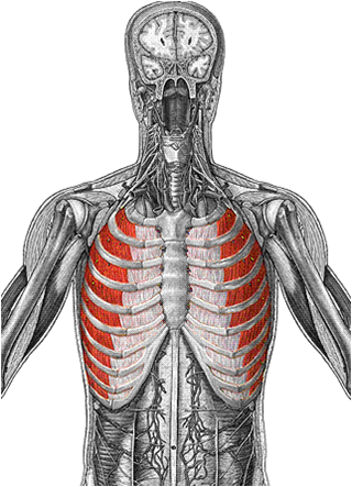
Anterior View of the Muscles of the Trunk | ClipArt ETC from etc.usf.edu (chest abdomen pelvis) slide # 1 (chest, abdomen, pelvis) carolyn kaut roth, rt (r)(mr)(ct)(m)(cv) fsmrt ceo imaging education associates www.imaginged.com candi@imaginged.com part i • planes of the chest, abdomen & pelvis • sectional anatomy of the chest & heart outline slide # 2 • mr imaging of the chest • sectional anatomy of the. Muscle anatomy drawing 12 photos of the muscle anatomy drawing anatomy muscle sketches, arm muscle anatomy drawing, back muscle anatomy drawing, human muscle anatomy drawing, muscle anatomy drawing, human muscles, anatomy muscle sketches, arm muscle anatomy drawing, back muscle anatomy drawing, human muscle anatomy. Signs and symptoms of pulled chest muscles. The fourth layer in the midregion is the rectus abdominis, which has vertically running muscle fibres that flex the trunk and stabilize the pelvis. One of the main smooth muscles inside the chest is the diaphragm. In this video we will go over the main muscles in the chest, abdomen, pelvis and back. Human muscle system, the muscles of the human body that work the skeletal system, that are under voluntary control, and that are concerned with movement. Deepest muscle of the abdomen, located deep to the internal oblique muscles;
Moving down the trunk of the cat from the chest to the abdomen, i was able to identify the latissimus dorsi, internal oblique, transverse abdominus, rectus abdominus, linea alba, and external oblique.
Chest muscles are responsible for adduction, internal rotation, and forwards flexion of the humerus. (chest abdomen pelvis) slide # 1 (chest, abdomen, pelvis) carolyn kaut roth, rt (r)(mr)(ct)(m)(cv) fsmrt ceo imaging education associates www.imaginged.com candi@imaginged.com part i • planes of the chest, abdomen & pelvis • sectional anatomy of the chest & heart outline slide # 2 • mr imaging of the chest • sectional anatomy of the. Angina is the term for chest pain caused by poor blood flow to the heart. Online quiz to learn muscles of the chest and abdomen; One of the most common symptoms of pulling a chest muscle is pain around the affected muscle. Muscles of the chest and abdomen— presentation transcript 24 muscles that move the arm (3 of 3) pectoralis major: Add to favorites 0 favs. Deepest muscle of the abdomen, located deep to the internal oblique muscles; Contracts with other abdominal muscles to compress the abdomen Related posts of muscles of the chest and abdomen muscle anatomy drawing. Abdominal muscle, any of the muscles of the anterolateral walls of the abdominal cavity, composed of three flat muscular sheets, from without inward: It arises from the fascia of the external oblique muscle. There are three muscular layers of the abdominal wall, with a fourth layer in the middle anterior region.
Muscles the dominant muscle in the upper chest is the pectoralis major. William blahd on webmd says that pulled muscle, strains, and tears can damage the muscle fibers and tendons. Chest muscles are responsible for adduction, internal rotation, and forwards flexion of the humerus. During a spasm, the muscle will feel stiff and tender if you apply pressure. A spasm may feel like a twitch or flutter and can occur with or without pain.
Anterior View of the Muscles of the Trunk | ClipArt ETC from etc.usf.edu A diaphragm spasm is an involuntary contraction of the muscle that divides the upper abdomen and chest. Muscles of the chest abdomen. Online quiz to learn muscles of the chest and abdomen; Between anterior chest and greater tubercle of humerus produces flexion at shoulder joint latissimus dorsi: Muscles the dominant muscle in the upper chest is the pectoralis major. Learn vocabulary, terms, and more with flashcards, games, and other study tools. Chest muscles function in respiration while abdominal muscles function in torso movement and in maintenance of balance and posture. (chest abdomen pelvis) slide # 1 (chest, abdomen, pelvis) carolyn kaut roth, rt (r)(mr)(ct)(m)(cv) fsmrt ceo imaging education associates www.imaginged.com candi@imaginged.com part i • planes of the chest, abdomen & pelvis • sectional anatomy of the chest & heart outline slide # 2 • mr imaging of the chest • sectional anatomy of the.
Rectus abdominis sternocleidomastoid trapezius serratus anterior linea alba rectus sheath pectoralis minor deltoid pectoralis major external oblique this problem has been solved!
Next to it on both sides of the body is. Muscles of the abdominal wall muscles of the abdominal wall. A heart attack results from blocked blood flow, often from a blood clot, to your heart muscle. Related posts of muscles of the chest and abdomen muscle anatomy drawing. There are three muscular layers of the abdominal wall, with a fourth layer in the middle anterior region. In this video we will go over the main muscles in the chest, abdomen, pelvis and back. The muscles of the abdomen were slightly less clear and. Abdominal muscle, any of the muscles of the anterolateral walls of the abdominal cavity, composed of three flat muscular sheets, from without inward: Muscles of the chest and abdomen learn by taking a quiz; Moving down the trunk of the cat from the chest to the abdomen, i was able to identify the latissimus dorsi, internal oblique, transverse abdominus, rectus abdominus, linea alba, and external oblique. Muscle anatomy drawing 12 photos of the muscle anatomy drawing anatomy muscle sketches, arm muscle anatomy drawing, back muscle anatomy drawing, human muscle anatomy drawing, muscle anatomy drawing, human muscles, anatomy muscle sketches, arm muscle anatomy drawing, back muscle anatomy drawing, human muscle anatomy. The fourth layer in the midregion is the rectus abdominis, which has vertically running muscle fibres that flex the trunk and stabilize the pelvis. The muscles on the anterior side of the forearm, such as the flexor carpi radialis and flexor.